Anatomy Of Chest Bone : Thoracic And Abdominal Muscles Lecturio Online Medical Library / These bones form by the thickening of a.
byTed Mcknight-
0
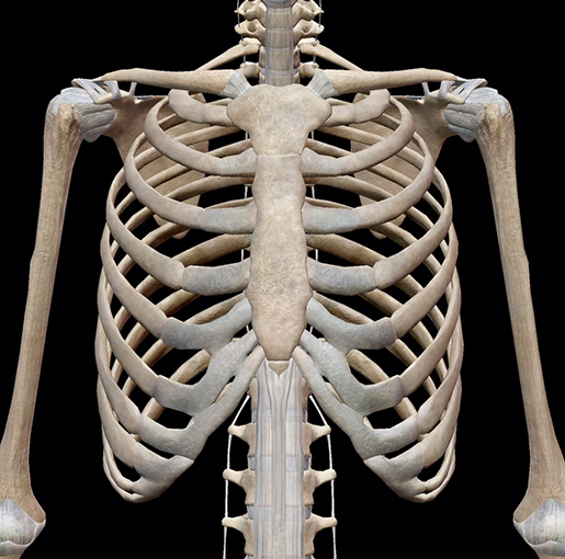
Anatomy Of Chest Bone : Thoracic And Abdominal Muscles Lecturio Online Medical Library / These bones form by the thickening of a.. Right upper anatomy is to physiology as geography is to history: Where is the sternum found. O bones—spine, ribs, clavicles, scapulae, humeri. Bones of the chest and upper back (posterior view). Chest bone, ribs, lung, heart, xiphoid process.
Sesamoid bones are generally small, flat and have an apex at one end. Different types of bones with differences are highlighted. Your rib cage, for example, acts like a shield around your chest to protect important organs inside such as your lungs and heart. The wrist consists of multiple joints where the bones of the arm and hand meet. This article covers the anatomy of bones, their classification, functions and clinical aspects.
3d Skeletal System Bones Of The Thoracic Cage from www.visiblebody.com The manubrium, sternal body, and xiphoid process. Sesamoid bones are generally small, flat and have an apex at one end. Learn about this topic at kenhub! Despite this it is easy to overlook important abnormalities of the bones which may be very subtle. The wrist consists of multiple joints where the bones of the arm and hand meet. In some patients an extra joint is seen in the anterior part of the first rib at the point where the bone meets the calcified cartilageneous part (arrow). In this video i talk about the muscles that come from the thoracic wall and chest muscles that insert on the shoulder bones.✅. The chest can be split into two parts;
It can help you understand our world more detailed and specific.
Originates/starts on the clavicle/collar bone and the sternum. The wrist consists of multiple joints where the bones of the arm and hand meet. The former is a type of connective tissue made up of cells suspended in a matrix: Where is the sternum found. Reference database of normal imaging from birth to age 16. These bones form by the thickening of a. Anatomy of the chest wall. Atlas of wrist mri anatomy. Bone basics and bone anatomy. Learn about this topic at kenhub! 12 photos of the anatomy bones chest. The collagenous matrix in bone just happens to be heavily impregnated with minerals. We hope you will use this picture in the study and helping chest and abdominal cavities with some organs removed.
Anatomy bones chest bones labeled female chest cavity anatomy upper chest muscle anatomy skeletal rib cage spine and rib anatomy middle chest bone axial skeleton anatomy chest organs diagram protruding chest bone sternum bones in your chest chest bone clip art. Bone also plays important roles in maintaining mineral homeostasis, as well as providing the environment for hematopoesis in marrow. 12 photos of the anatomy bones chest. Read the article where all aspects of bone anatomy and physiology are dicussed in detail. The skull is a bony structure that supports the face and forms a protective cavity for the brain.
Ribs Sternum from wp.stu.ca Originates/starts on the clavicle/collar bone and the sternum. Human chest bone structure parts of the chest bones. Bone basics and bone anatomy. It originates at your clavicle, ribs, and sternum, and inserts into the upper portion of your humerus (upper arm bone from elbow to shoulder.) The former is a type of connective tissue made up of cells suspended in a matrix: Bones of the chest and upper back (posterior view). Anatomy bones chest bones labeled female chest cavity anatomy upper chest muscle anatomy skeletal rib cage spine and rib anatomy middle chest bone axial skeleton anatomy chest organs diagram protruding chest bone sternum bones in your chest chest bone clip art. Despite this it is easy to overlook important abnormalities of the bones which may be very subtle.
Long bones are categorised by their tubular shaft (diaphysis) with a rounded end (epiphysis) on each end.
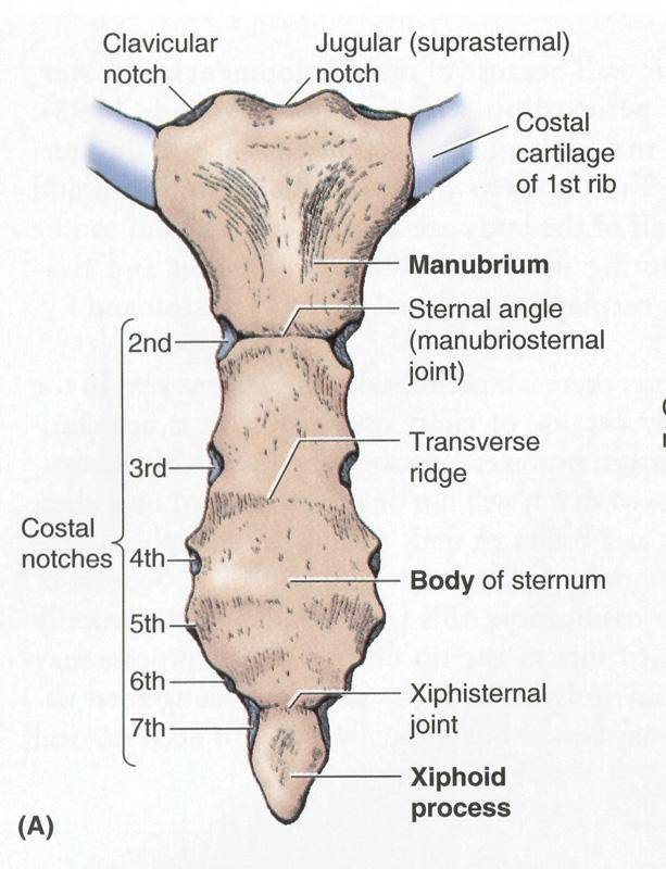
When a patient flexes the neck forward, the prominent process is usually that of the 7th cervical. Bones support and protect the body and its organs. Atlas of anatomy of the human body: It originates at your clavicle, ribs, and sternum, and inserts into the upper portion of your humerus (upper arm bone from elbow to shoulder.) The medial anterior chest is defined by the sternum, which consists of 3 flat polygonal bones: Anatomists talk about both bone and bones. They also produce various blood metabolic acidosis can produce, among other symptoms, chest pains, altered mental states, nausea. Inserts/attaches on the humerus/upper arm. Where is the sternum found. Anatomy is the amazing science. Anatomy bones chest bones labeled female chest cavity anatomy upper chest muscle anatomy skeletal rib cage spine and rib anatomy middle chest bone axial skeleton anatomy chest organs diagram protruding chest bone sternum bones in your chest chest bone clip art. Pathology of the heart, mediastinum, lungs and pleura. Bones of the chest and upper back (posterior view).
Pathology of the heart, mediastinum, lungs and pleura. Learn about this topic at kenhub! Bone of chest and their parts. Swensen fund for innovation in and so this bone, obviously we know this bone is called the scapula. Sesamoid bones are generally small, flat and have an apex at one end.
Chest Bone Ribs Lung Heart Xiphoid Process Sternum Anatomy from www.anatomynote.com The skull is a bony structure that supports the face and forms a protective cavity for the brain. The former is a type of connective tissue made up of cells suspended in a matrix: Anatomy is the amazing science. We hope you will use this picture in the study and helping chest and abdominal cavities with some organs removed. It describes the theatre of events. Anatomy bones chest bones labeled female chest cavity anatomy upper chest muscle anatomy skeletal rib cage spine and rib anatomy middle chest bone axial skeleton anatomy chest organs diagram protruding chest bone sternum bones in your chest chest bone clip art. Atlas of wrist mri anatomy. These bones form by the thickening of a.
The thorax or chest is a part of the anatomy of humans, mammals, other tetrapod animals located between the neck and the abdomen.
The pectoralis major and minor. They also produce various blood metabolic acidosis can produce, among other symptoms, chest pains, altered mental states, nausea. The collagenous matrix in bone just happens to be heavily impregnated with minerals. Language and terminology for the study of the anatomy of the thorax. Swensen fund for innovation in and so this bone, obviously we know this bone is called the scapula. All of the anatomical and important histological facts about the bones, together with the clinical relations, are going to be desrcibed in this article. They are always longer than they are wide the vertebrae are irregular bones. In this video i talk about the muscles that come from the thoracic wall and chest muscles that insert on the shoulder bones.✅. Atlas of anatomy of the human body: Flat bones form by membranous bone formation, whereas long bones are formed by a combination of endochondral and membranous bone formation. Have you ever seen fossil remains of dinosaur and ancient human bones in textbooks, television, or in person at a museum? Long bones are categorised by their tubular shaft (diaphysis) with a rounded end (epiphysis) on each end. This anatomical midline can be useful in assessing for symmetry in breast augmentation or in performing a median sternotomy.
It originates at your clavicle, ribs, and sternum, and inserts into the upper portion of your humerus (upper arm bone from elbow to shoulder) anatomy of chest. And we want to know some borders about it.